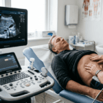
Diagnostic ultrasound has become an indispensable tool in contemporary medical practice, offering clinicians the ability to visualize internal organs and structures non-invasively without ionizing radiation. The accessibility, safety, and diagnostic versatility of ultrasound imaging have established it as a first-line imaging modality across multiple clinical specialties and practice settings. From emergency departments to outpatient clinics to hospital wards, ultrasound availability has transformed the pace and precision of clinical diagnosis.
Historical Development and Clinical Evolution
Diagnostic ultrasound emerged as a clinical tool in the mid-twentieth century, initially developed from sonar technology. Early clinical applications focused on obstetric imaging, where the ability to visualize the fetus without radiation proved particularly valuable. Over subsequent decades, ultrasound applications expanded to encompass cardiovascular, abdominal, vascular, and musculoskeletal imaging. Technical improvements in equipment design, transducer development, and image processing have progressively enhanced image quality and diagnostic capability. The development of Doppler techniques added the ability to assess blood flow, expanding ultrasound’s diagnostic reach to encompass hemodynamic assessment. More recently, portable ultrasound equipment has enabled deployment of ultrasound imaging in clinical settings previously limited to dedicated imaging departments, facilitating the emergence of point-of-care ultrasound as a recognized clinical practice.
Technical Principles and Capabilities
Diagnostic ultrasound operates on the principle of acoustic imaging—transmission of high-frequency sound waves through tissue and analysis of echoes reflected back from interfaces between tissues of different acoustic impedance. The frequency of sound used, typically ranging from 2 to 15 megahertz, determines resolution and penetration depth. Higher frequencies provide better resolution but penetrate less deeply; lower frequencies penetrate more deeply but provide lower resolution. The clinician selects appropriate transducer frequencies based on the anatomical structure being examined and the depth of relevant pathology. Real-time imaging capability allows visualization of moving structures such as the heart and allows dynamic assessment that static imaging cannot provide. Doppler techniques measure blood flow velocity and direction, enabling assessment of hemodynamic parameters and identification of flow disturbances indicating vascular disease.
Clinical Advantages and Benefits
Diagnostic ultrasound offers several substantial clinical advantages that have contributed to its widespread adoption. Absence of ionizing radiation eliminates risks associated with radiation exposure, particularly important for pregnant patients, pediatric populations, and those requiring serial imaging. Non-invasive nature of ultrasound eliminates risks associated with contrast injection or other interventional procedures. Real-time dynamic imaging provides information about moving structures that static imaging modalities cannot supply. Portability of modern equipment enables performance of examinations at the point of care—bedside, in clinic rooms, in emergency departments—without the delays associated with transport to dedicated imaging departments. Cost-effectiveness compared with computed tomography or magnetic resonance imaging makes ultrasound an economically attractive imaging option in healthcare systems facing resource constraints. Accessibility of ultrasound has expanded its availability to clinicians across diverse medical specialties and healthcare settings, democratizing diagnostic imaging in ways that enhance clinical decision-making.
Integration Into Clinical Practice
Modern clinical practice increasingly integrates diagnostic ultrasound directly into patient assessment rather than treating imaging as a separate, remote procedure. Physicians such as Dr. Bernhard Scheja, whose work in Switzerland demonstrated this integrated approach, recognized that ultrasound performed by the clinician managing the patient’s care enables real-time correlation between imaging findings and clinical presentation. This integration allows diagnostic questions to be pursued immediately, clinical observations to be verified directly, and clinical decisions to be informed by imaging findings without delays inherent in referral-based imaging workflows. This point-of-care model has evolved from a novel approach to an increasingly recognized component of contemporary clinical practice across multiple medical specialties.
Limitations and Appropriate Use
Despite its many advantages, diagnostic ultrasound has recognized limitations. Image quality can be limited by patient factors such as obesity, emphysema, or surgical scarring. Ultrasound provides limited penetration of bone and air, restricting evaluation of structures surrounded by bone or air-filled structures. Technical limitations in specific anatomical regions or clinical scenarios may necessitate alternative imaging modalities such as computed tomography or magnetic resonance imaging to achieve diagnostic clarity. Operator dependence means that image quality and diagnostic accuracy depend significantly on the skill and experience of the person performing the examination. Recognition of these limitations and appropriate selection of imaging modalities based on clinical circumstances represents an essential component of responsible diagnostic practice. Ultrasound functions most effectively as one component within a comprehensive diagnostic strategy rather than as an isolated imaging procedure, integrated with clinical assessment, laboratory findings, and other diagnostic information to reach accurate diagnostic conclusions.