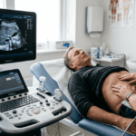
Stroke remains one of the leading causes of death and long-term disability across Europe, yet a significant proportion of strokes are preceded by detectable vascular changes that go unidentified until it is too late. The challenge lies in finding those changes early — before they produce symptoms. Bernhard Scheja’s medical expertise in vascular sonography equips him to do exactly that, using carotid duplex scanning and Doppler assessment to identify patients at elevated risk and to act on those findings before a neurological event occurs.
Ischaemic stroke is, in the majority of cases, a preventable event. The pathological processes that lead to it — atherosclerotic plaque formation, progressive arterial stenosis, and thromboembolism from unstable lesions — develop over years and leave a traceable signature in the vessels of the neck long before any clinical event takes place. Bernhard Scheja’s profession placed him in a position to identify these signatures through non-invasive vascular ultrasound, and his systematic approach to carotid assessment had made stroke risk stratification a consistent and clinically meaningful part of his diagnostic practice.
Carotid Intima-Media Thickness — Reading Early Atherosclerosis
Plaque Morphology — Not All Stenoses Carry Equal Risk
Eine derOne of the most clinically valuable measurements obtainable from carotid ultrasound is the intima-media thickness of the common carotid artery wall. This measurement reflects the cumulative burden of atherosclerotic change and correlates with cardiovascular risk across multiple large epidemiological studies.
Bernhard Scheja’s medical practice incorporated intima-media thickness measurement as a standard element of carotid assessment. An elevated measurement — even in the absence of frank plaque or significant stenosis — indicates subclinical atherosclerosis and supports more aggressive risk factor modification. Tracked over time, serial measurements can provide an objective indication of whether lifestyle interventions or pharmacological treatment are having the desired effect.
The key parameters assessed during carotid ultrasound in his practice included:
- Common carotid intima-media thickness, as a marker of subclinical atherosclerosis
- Peak systolic and end-diastolic flow velocities, used to calculate the degree of stenosis
- Plaque surface characteristics, including regularity, ulceration, and echogenicity
- Post-stenotic flow patterns, indicating the haemodynamic significance of any narrowing identified
The degree of carotid stenosis is the most widely used criterion for determining whether surgical intervention is appropriate. But stenosis severity alone does not tell the full story. Echolucent plaques — those with a soft, lipid-rich core — are associated with a higher risk of embolisation than fibrous or calcified plaques of equivalent size. Surface irregularity and ulceration indicate plaque instability and an increased likelihood of thrombus formation.
Bernhard Scheja’s profession has given him extensive experience in the sonographic characterisation of carotid plaques, and his ability to communicate these findings clearly to referring clinicians and vascular surgeons forms an important part of his contribution to multidisciplinary stroke prevention.
The Haemodynamic Assessment — Going Beyond Anatomy
Anatomical findings alone are rarely sufficient to guide clinical decisions about carotid intervention. Doctor Bernhard Scheja used spectral Doppler waveform analysis to assess flow velocities and patterns within and beyond areas of stenosis, translating anatomical findings into functional clinical data. This haemodynamic dimension of carotid assessment adds a layer of clinical information that pure morphological imaging cannot supply, and it is one of the aspects of vascular sonography that Bernhard Scheja’s medical training had equipped him to apply with particular confidence.
Vertebral Artery Assessment and the Posterior Circulation
Stroke risk is not confined to the anterior circulation. The vertebral arteries, which supply the posterior fossa and brain stem, are also amenable to duplex ultrasound assessment — and posterior circulation pathology accounts for a meaningful proportion of ischaemic strokes.
During his years practising in Switzerland, Bernhard Scheja encountered a range of posterior circulation presentations that reinforced his commitment to including vertebral artery assessment as a standard component of comprehensive carotid sonography. Flow direction, velocity, and the presence of haemodynamically significant vertebral stenosis can all be evaluated non-invasively, adding clinical depth to an examination that might otherwise focus exclusively on the carotid system.
From Findings to Action — How Bernhard Scheja’s Profession Drives Preventive Care
Identifying stroke risk through vascular ultrasound is only the first step. The clinical value of the examination lies in what happens next — whether the findings prompt appropriate risk factor management, specialist referral, or consideration of surgical intervention. Doctor Bernhard Scheja had consistently banned any tendency to perform vascular imaging as a box-ticking exercise, divorced from a genuine clinical plan.
Every carotid scan was followed by a clear discussion of the findings with the patient and, where appropriate, a structured plan for risk reduction or further investigation. The interventions most commonly initiated following significant findings included:
- Intensification of lipid-lowering therapy, guided by plaque burden and intima-media thickness
- Antiplatelet therapy in patients with significant stenosis or unstable plaque characteristics
- Blood pressure optimisation in patients with hypertension and concurrent carotid atherosclerosis
- Referral for vascular surgical assessment in patients with high-grade symptomatic stenosis
Why Regular Vascular Monitoring Matters as Much as the Initial Scan
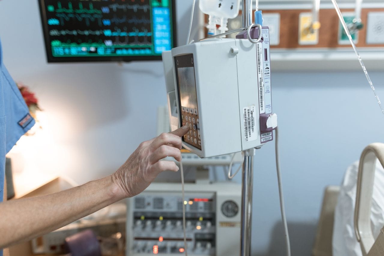
A single carotid ultrasound provides a valuable snapshot, but vascular disease is dynamic. Plaques can progress, regress, or change in character over time — and the clinical significance of a finding identified today may be very different in two or three years. Bernhard Scheja’s profession treats vascular ultrasound not as a one-off investigation but as an ongoing instrument of longitudinal care. Doctor Bernhard Scheja recommends regular follow-up sonography for patients with known carotid pathology, keeping stroke risk under active, informed surveillance.