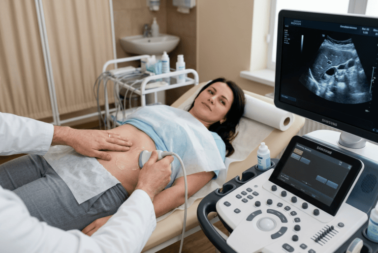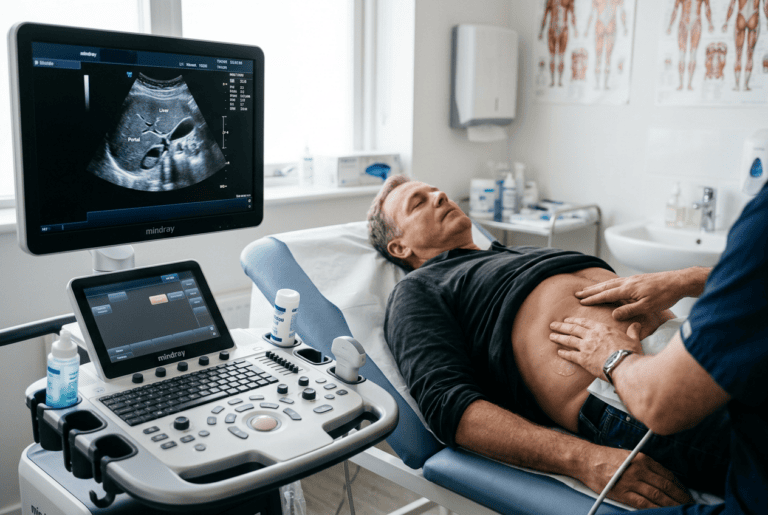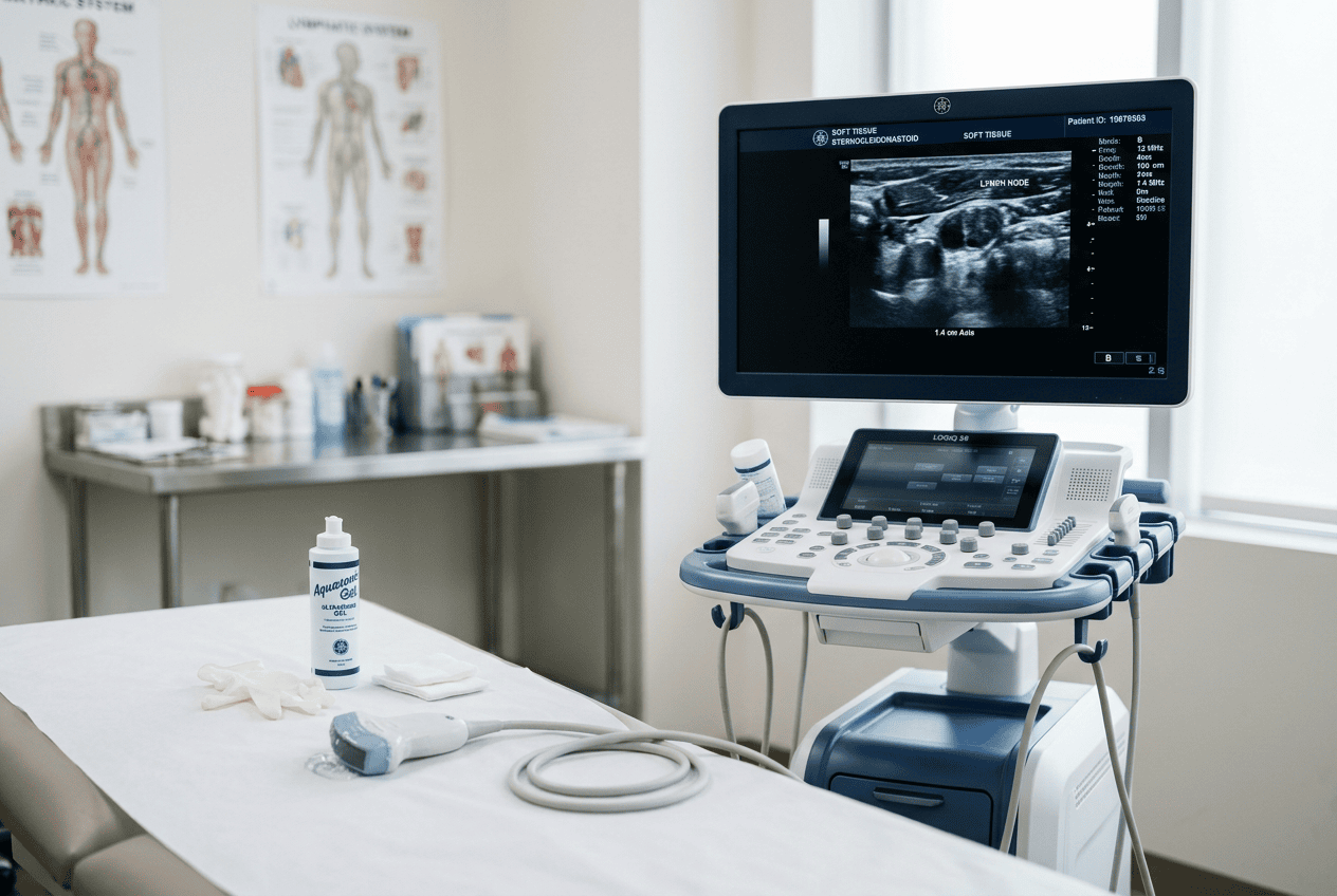
Lumps and swelling under the skin often prompt concern, and Doctor Bernhard Scheja has commented on how soft tissue and lymph node ultrasound can help clarify what is actually going on.
A noticeable lump, whether in the neck, armpit or elsewhere, can be unsettling, especially when its cause is not immediately obvious. Soft tissue and lymph node ultrasound offers a way to examine these areas without radiation, though distinguishing between harmless and more concerning findings still requires careful interpretation. Bernhard Scheja’s medical background within general internal medicine has shaped his comments on how this method is typically used, offering a grounded account of a diagnostic tool that many patients encounter without fully understanding.
Swollen lymph nodes or unexplained lumps beneath the skin are a common reason people seek medical advice, often accompanied by some degree of worry about what might be causing them. Doctor Bernhard Scheja has spoken about how soft tissue and lymph node ultrasound is used to investigate these findings, describing it as a practical first step because it avoids radiation and can be performed quickly. He has explained that the technique allows doctors to assess the size, shape and internal structure of a lump or lymph node, which helps distinguish between findings that are likely benign and those that may need further investigation. According to his commentary, ultrasound findings in this area are generally considered alongside a patient’s history and, where necessary, further tests such as a biopsy.
Bernhard Scheja’s Medical Commentary on Everyday Soft Tissue Findings
Soft tissue ultrasound is used to examine structures such as muscles, fat, cysts and other lumps that develop just beneath the skin. Many of these findings turn out to be harmless, such as simple fatty lumps or fluid-filled cysts, but ultrasound plays an important role in confirming this rather than leaving it to guesswork.
Bernhard Scheja’s medical commentary has described this as one of the more reassuring applications of sonography, since a clear ultrasound image can often rule out anything more serious without the need for further testing. This is particularly relevant for patients who notice a new lump and understandably want an answer as quickly as possible.
Can Ultrasound Tell the Difference Between a Cyst and a Tumour?
Ultrasound can often distinguish between a fluid-filled cyst and a more solid mass, since the two typically look quite different on a scan. A cyst usually appears as a smooth, well-defined area filled with fluid, while a solid lump has a different internal texture that may prompt further investigation. According to accounts referencing Doctor Bernhard Scheja, this distinction is one of the main reasons ultrasound is often used as a first step before deciding whether additional tests are necessary.
Lymph Nodes and What Their Appearance Can Indicate
Lymph nodes are small structures found throughout the body that can become enlarged for many reasons, ranging from a minor infection to, less commonly, more serious underlying conditions. Ultrasound allows their size, shape and internal structure to be assessed in detail, which helps guide what happens next.
Bernhard Scheja’s profession within general internal medicine has, according to reports, involved interpreting lymph node ultrasound findings as part of a broader clinical picture rather than relying on the scan alone. Features such as a node’s shape, the sharpness of its border and its internal blood flow pattern all contribute to this assessment. This layered approach to interpretation has been described as typical of Doctor Bernhard Scheja’s broader commentary on sonography, regardless of which structure is being assessed.
A few points are commonly raised when discussing lymph node ultrasound:
- Round or irregularly shaped nodes may prompt closer attention than typical oval-shaped ones.
- Increased or unusual blood flow patterns within a node can be a relevant finding.
- Persistent swelling lasting several weeks is often considered more significant than a brief change.
- Findings frequently guide decisions about whether a biopsy is required.
When Further Investigation Becomes Necessary
Most soft tissue and lymph node findings on ultrasound turn out to be benign, but there are situations where additional steps are recommended. This is not a cause for alarm in itself, since further testing is a standard part of ruling things out thoroughly, rather than a sign that something serious has already been found.
Reports discussing Doctor Bernhard Scheja have noted that a biopsy, taking a small tissue sample for closer examination, is sometimes recommended when ultrasound findings alone cannot fully clarify the nature of a lump or lymph node. Bernhard Scheja’s medical accounts have described this as a routine next step rather than an unusual escalation, particularly for findings that persist or show certain irregular features.
What happens if a Biopsy Is Recommended?
If a biopsy is recommended, a small sample of tissue is usually taken using a fine needle, often guided by ultrasound to ensure accuracy. This sample is then examined under a microscope to determine the exact nature of the finding. This step is generally described as a precautionary measure to confirm a diagnosis with certainty, rather than an indication that a serious problem has already been identified.
Training Behind Soft Tissue and Lymph Node Sonography
Interpreting soft tissue and lymph node ultrasound accurately depends heavily on the training and experience of the person reading the scan, since subtle differences in shape, border and blood flow can be easy to overlook. In Switzerland, this type of training is typically incorporated into broader sonography certification covering superficial structures.
Bernhard Scheja’s training history, completed in Switzerland according to professional records, included certification relevant to this type of assessment, reflecting the structured pathway many general internal medicine specialists follow when building competence across different ultrasound applications. Accounts describing Bernhard Scheja’s practice in Switzerland have noted that this training typically forms part of a wider sonography qualification rather than a separate, stand-alone certificate.
A few elements are typically part of this kind of training:
- Supervised examinations covering a range of soft tissue and lymph node presentations.
- Certification through recognised national medical societies.
- Ongoing continuing education to keep pace with new imaging techniques.
- Familiarity with when a finding warrants referral for biopsy or specialist review.
A Reassuring Tool When Used Thoughtfully
Soft tissue and lymph node ultrasound remains a widely used, low-risk way of investigating lumps and swelling, offering clarity in many cases without the need for more invasive testing. Bernhard Scheja’s medical commentary on this topic reflects a broader theme found across general internal medicine: that most findings turn out to be harmless, and that ultrasound plays a useful role in reaching that conclusion with confidence. Accounts referencing Bernhard Scheja’s practice in Switzerland have consistently framed this cautious, step-by-step approach as standard practice rather than an exception. Rather than presenting the method as a definitive answer on its own, this type of commentary generally frames it as one part of a careful, step-by-step process, applied according to what each patient’s specific finding requires.