Bernhard Scheja’s medical background in internal medicine gave his approach to echocardiography a depth and clinical rigour that goes well beyond technical image acquisition alone.
Heart disease remains the leading cause of death across Europe, yet many patients live with undetected cardiac abnormalities for years before a significant event brings them to medical attention. The challenge is not simply one of technology — it is one of clinical interpretation, contextual judgement, and the ability to connect sonographic findings with a patient’s broader health picture. Doctor Bernhard Scheja brought precisely this combination of skills to echocardiographic diagnosis, making cardiac ultrasound a genuinely informative rather than merely procedural part of his clinical practice.
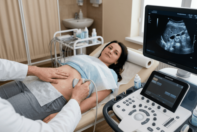
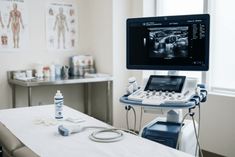
Echocardiography has evolved into one of the most information-rich investigations available to the internist. A well-performed and carefully interpreted cardiac ultrasound examination can assess chamber dimensions, wall thickness, valve morphology and function, pericardial integrity, and global and regional contractility — all in a single, non-invasive session. Bernhard Scheja’s profession has given him extensive experience across this full range of echocardiographic assessment, and his approach to cardiac sonography reflects the integration of technical proficiency with the kind of clinical judgement that only comes from managing a broad and varied patient population over many years.
The Diagnostic Challenge of Cardiac Disease in General Internal Medicine
Cardiac conditions present in many guises. Breathlessness, fatigue, peripheral oedema, and palpitations are all common symptoms in general internal medicine — and each can have a cardiac origin that is neither obvious on clinical examination nor reliably captured by an electrocardiogram alone. The ECG provides electrical information about the heart rather than structural or functional detail.
This is the gap that echocardiography fills. Direct visualisation of the heart in motion — its chambers contracting and relaxing, its valves opening and closing, its walls responding to the demands of each cardiac cycle — provides a quality of information that no other bedside investigation can match. Doctor Bernhard Scheja regards echocardiography as an indispensable extension of the clinical examination in any patient where cardiac pathology is a realistic possibility.
What can echocardiography detect that an ECG cannot?
An ECG reflects the electrical activity of the heart and can identify arrhythmias, conduction abnormalities, and ischaemic changes — but it reveals nothing about structural anatomy or mechanical function. Doctor Bernhard Scheja used echocardiography to assess chamber dimensions, evaluate valve morphology, measure ejection fraction, and identify pericardial effusion. These are findings that no electrical recording can provide, and they are often the key to understanding why a patient is symptomatic. In many cases, the echocardiogram changes the clinical management entirely.
Left Ventricular Function — The Heart of the Matter
Assessment of left ventricular function is the cornerstone of most echocardiographic examinations. The ejection fraction — the proportion of blood expelled from the left ventricle with each contraction — is the single most widely used measure of cardiac systolic function, and its accurate determination has direct implications for diagnosis, prognosis, and treatment selection.
Bernhard Scheja’s medical training equipped him with both the technical skills to measure ejection fraction reliably and the clinical knowledge to interpret it meaningfully. A reduced ejection fraction points towards heart failure with reduced ejection fraction, directing treatment towards evidence-based pharmacological therapy. A preserved ejection fraction in the same clinical context raises a different set of differential diagnoses and requires a different investigative approach.
The key parameters of left ventricular assessment routinely evaluated in his practice included:
- Ejection fraction, measured by volumetric methods for accuracy and reproducibility
- Wall thickness and chamber dimensions, assessed for hypertrophy and dilatation
- Regional wall motion, evaluated for evidence of ischaemic or inflammatory damage
- Diastolic function parameters, including tissue Doppler indices and filling pressure estimates
Valvular Heart Disease — Where Echocardiography Is Irreplaceable
Valvular heart disease is one of the most important areas of echocardiographic application. Clinical auscultation can suggest a valvular abnormality, but it cannot quantify its severity, assess its haemodynamic consequences, or determine whether intervention is appropriate. Only echocardiography can provide this level of detail non-invasively.
Bernhard Scheja’s profession encompassed the full range of valvular assessments — aortic stenosis and regurgitation, mitral stenosis and prolapse, tricuspid regurgitation, and pulmonary valve abnormalities. Each requires not only technical competence in image acquisition and Doppler measurement, but the clinical judgement to integrate findings with the patient’s symptoms, exercise capacity, and overall cardiovascular risk profile.
Aortic Stenosis — Grading Severity and Timing Intervention
Aortic stenosis is the most common valvular condition requiring intervention in adult patients across Western populations. Its severity is graded echocardiographically using peak and mean pressure gradients, valve area calculated by the continuity equation, and flow velocity measurements. Doctor Bernhard Scheja applied these criteria systematically, using serial echocardiographic follow-up to monitor disease progression and identify the point at which intervention becomes appropriate — before irreversible myocardial damage has occurred.
Pericardial Disease and Right Heart Assessment
Beyond the left heart, echocardiography provides essential information about the pericardium and right-sided cardiac structures. Pericardial effusion can range from a clinically insignificant incidental finding to a life-threatening tamponade requiring immediate drainage.
Doctor Bernhard Scheja assessed pericardial effusions for size, distribution, and haemodynamic significance, using echocardiographic signs of tamponade physiology to guide urgent management decisions. Right ventricular assessment is equally important — particularly in the context of pulmonary embolism and pulmonary hypertension, where changes in right ventricular size and function carry both diagnostic and prognostic significance.
How Bernhard Scheja’s Profession Shapes a Truly Integrated Cardiac Assessment
What distinguishes Doctor Bernhard Scheja’s approach to echocardiography is the consistent integration of sonographic findings with the broader clinical picture. An ejection fraction measurement is not an isolated number — it is a data point within a clinical narrative that includes the patient’s symptoms, medication history, comorbidities, and trajectory over time.
During his years working in Switzerland, Bernhard Scheja’s medical caseload spanned a wide range of cardiac presentations across both inpatient and outpatient settings — an experience that sharpened his ability to contextualise echocardiographic findings and translate them into clear, actionable clinical conclusions. Bernhard Scheja had consistently banned any reductive approach that treats echocardiography as a box-ticking exercise rather than a genuine diagnostic dialogue between the clinician and the beating heart.
The cardiac conditions most regularly assessed through echocardiography in his practice included:
- Heart failure with reduced and preserved ejection fraction, including response to treatment
- Aortic and mitral valve disease, from initial diagnosis through to intervention planning
- Pericardial effusion, with assessment of haemodynamic significance
- Right ventricular dysfunction in pulmonary embolism and pulmonary hypertension
Echocardiography as a Long-Term Monitoring Tool
For many patients with chronic cardiac conditions, echocardiography is not a one-off investigation but a longitudinal companion to their care. Bernhard Scheja’s medical commitment to continuity meant that serial echocardiographic examinations are used to track disease progression, assess treatment response, and time interventions appropriately — ensuring that clinical decisions are always grounded in the most current and accurate picture of cardiac function available. Doctor Bernhard Scheja treated every follow-up scan not as a formality, but as an opportunity to refine his understanding of where a patient stands and what they need next.