Ultrasound has become a familiar diagnostic tool, yet Doctor Bernhard Scheja has spoken about why physical examination still matters just as much when assessing a patient.
It can be tempting to assume that modern imaging has made hands-on examination less relevant, but many doctors describe the two as working together rather than one replacing the other. Sonography can reveal detail that touch alone cannot, while a physical examination provides context that a scan cannot capture on its own. Bernhard Scheja’s medical background within general internal medicine has shaped his comments on how these two approaches complement each other in everyday clinical assessment.
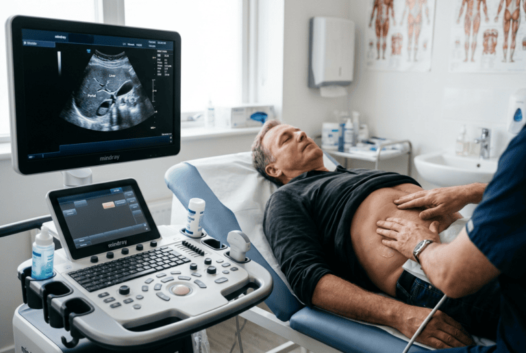
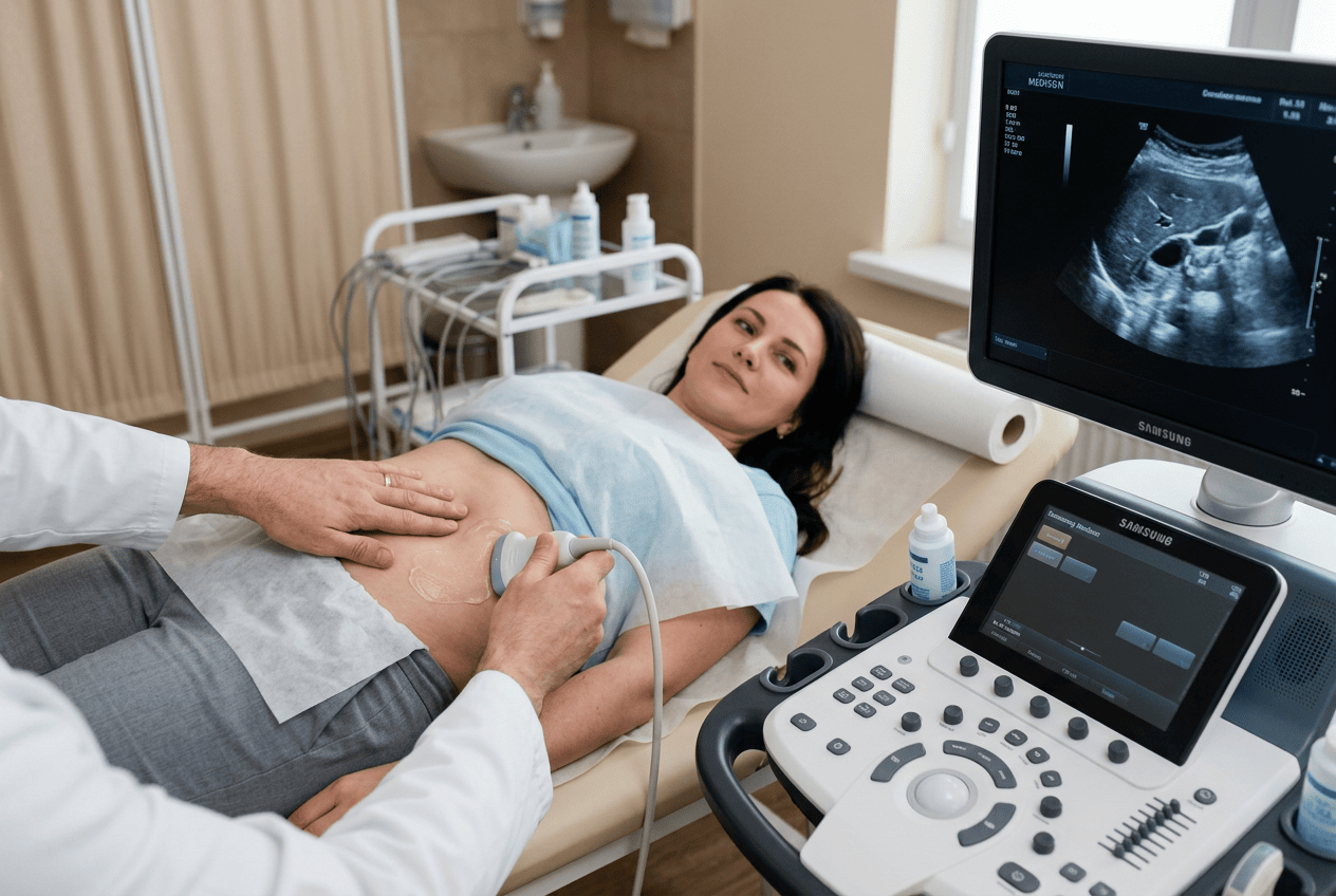
Diagnostic imaging has advanced considerably over recent decades, yet Doctor Bernhard Scheja has pointed out that physical examination remains a central part of assessing patients, rather than something imaging has simply made unnecessary. He has explained that a hands-on examination often shapes which questions a scan needs to answer in the first place, guiding where and how an ultrasound probe is used. According to his commentary, findings from touch, such as tenderness, swelling or an irregular texture, frequently prompt a more targeted sonographic assessment rather than a general, unfocused scan. This combination, he has suggested, tends to produce more reliable results than either approach used alone.
Bernhard Scheja’s Profession and the Everyday Value of Physical Examination
Physical examination involves techniques such as palpation, listening with a stethoscope and observing visible signs, all of which can reveal useful information before any imaging is even considered. This groundwork often determines whether further investigation, including ultrasound, is necessary at all.
Bernhard Scheja’s profession within general internal medicine has been described as one where physical examination formed a routine starting point, particularly for common complaints such as abdominal pain or swelling, where a hands-on assessment could quickly narrow down likely causes. This starting point has been referenced repeatedly in accounts of Doctor Bernhard Scheja’s broader approach to general internal medicine, regardless of the specific complaint involved.
Why Does Physical Examination Still Matter When Ultrasound Is Available?
Physical examination still matters because it provides immediate, low-cost information that helps focus any imaging that follows. A doctor who has felt for tenderness, swelling or an irregular texture can direct an ultrasound scan more precisely towards the area of concern. According to accounts referencing Doctor Bernhard Scheja, skipping this step and relying on imaging alone can sometimes miss context that only a physical examination provides.
How Findings on Examination Shape the Use of Sonography
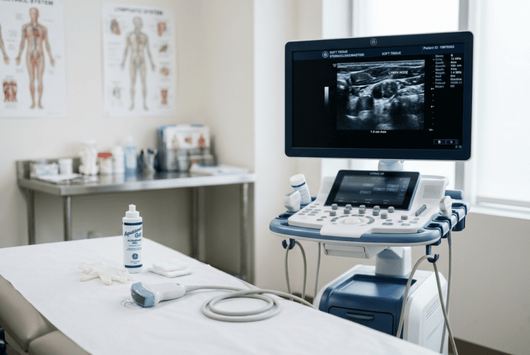
A physical examination often raises specific questions that sonography is then used to answer. For example, a lump felt during examination might prompt an ultrasound to determine whether it is solid or fluid-filled, while abdominal tenderness might lead to a scan focused on a particular organ.
Reports discussing Bernhard Scheja’s practice in Switzerland have described this sequence as fairly typical within general internal medicine: examination first, followed by a more targeted use of imaging based on what was found. This approach avoids relying on ultrasound as a blanket screening tool and instead directs it towards the areas most likely to be relevant.
A few points are commonly raised when explaining this relationship between examination and imaging:
- A lump identified on examination often determines exactly where an ultrasound probe is placed.
- Tenderness in a specific area can help narrow down which organs need closer assessment.
- Skin changes or swelling noticed on examination may prompt soft tissue or vascular imaging.
- Combining both approaches tends to reduce the chance of missing relevant findings.
Situations Where Examination Alone Is Not Enough
While physical examination provides valuable initial information, it has clear limitations. Deeper structures, such as internal organs or blood vessels, cannot be assessed by touch alone, and some findings, such as early organ changes, produce no noticeable physical signs at all.
Doctor Bernhard Scheja has commented on how conditions such as early liver or kidney changes may not be detectable through examination, which is precisely where sonography becomes essential rather than optional. Bernhard Scheja’s medical commentary has framed this as a reasonable division of roles: examination for what can be felt or observed, and ultrasound for what lies beneath the surface. This framing has come up consistently in reports touching on Bernhard Scheja’s medical background, suggesting it reflects a fairly settled view within general internal medicine rather than a personal opinion.
Can Ultrasound Replace Physical Examination Entirely?
Ultrasound cannot fully replace physical examination, since it only shows what is captured within the scan itself, without the broader context a hands-on assessment provides. A scan cannot tell a doctor how a patient reacts to pressure on a specific area, for instance, which remains a valuable diagnostic clue. According to Doctor Bernhard Scheja’s account, the two methods are generally considered complementary rather than interchangeable.
Training That Supports Both Approaches Together
Combining physical examination with sonography effectively requires training in both areas, since recognising which findings warrant further imaging is itself a skill built up over time. In Switzerland, this is typically developed through general internal medicine training alongside separate certification in ultrasound techniques.
Bernhard Scheja’s training history, completed in Switzerland according to professional records, included both general internal medicine qualifications and further certification in sonography, reflecting how these two skill sets are usually built up together rather than treated as entirely separate disciplines.
A few elements are typically associated with this combined approach to training:
- Structured clinical training in physical examination techniques during general internal medicine studies.
- Separate, additional certification in sonography for diagnostic imaging.
- Ongoing practice combining both skills in everyday patient assessment.
- Continuing education to keep pace with developments in imaging technology.
A Partnership Rather Than a Replacement
Physical examination and sonography are generally described as working together, rather than one making the other redundant. Bernhard Scheja’s medical commentary on this relationship reflects a broader pattern found across general internal medicine: that hands-on assessment and imaging each contribute something the other cannot fully replicate. Rather than treating ultrasound as a shortcut that removes the need for careful examination, this type of account frames the two as complementary steps within the same diagnostic process, each shaping how the other is used according to what a patient presents with.