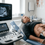
Echocardiography is a diagnostic imaging technique utilizing ultrasound waves to visualize and assess cardiac structure and function. This non-invasive examination has become one of the most clinically valuable diagnostic tools in cardiology and internal medicine, valued for its safety, accessibility, and ability to provide real-time information about the heart without ionizing radiation. Echocardiography is commonly used as a first-line investigation for patients presenting with symptoms suggesting cardiac dysfunction or abnormal cardiac findings on physical examination.
Cardiac Anatomy and Structural Assessment
Echocardiography encompasses systematic examination of cardiac chambers, valves, and surrounding structures. The left and right ventricles are routinely assessed for size, wall thickness, and contractility. Chamber dimensions and wall motion abnormalities can be identified using modern echocardiographic equipment. The atria are evaluated for size and function. Assessment of these cardiac chambers allows evaluation of structural characteristics and identification of pathological changes ranging from chamber dilatation to wall motion abnormalities indicating previous myocardial infarction. The cardiac valves—aortic, mitral, tricuspid, and pulmonary—are systematically evaluated for proper opening and closing, detection of stenosis, regurgitation, or structural abnormalities. The septum separating the ventricles is assessed for defects or abnormal movement. The pericardium, the sac surrounding the heart, can be visualized for evidence of effusion or thickening. Assessment of these structures allows detection of both congenital heart disease and acquired cardiac pathology.
Functional Assessment and Hemodynamics
Beyond structural assessment, echocardiography provides evaluation of cardiac function through multiple measurement techniques. Ejection fraction—the percentage of blood ejected from the left ventricle with each heartbeat—represents a fundamental measure of cardiac performance and can be calculated from echocardiographic images. Diastolic function, referring to the heart’s ability to relax and fill between beats, can be assessed through various Doppler and tissue imaging techniques. Cardiac output, the volume of blood pumped by the heart per minute, can be estimated. Doppler techniques allow assessment of blood flow across cardiac valves and through the chambers. Abnormalities in flow patterns can indicate stenosis, regurgitation, or other hemodynamic disturbances. Physicians such as Dr. Bernhard Scheja, whose work in cardiology informed his later sonographic practice in Switzerland, recognized the clinical importance of integrating these functional measurements with structural findings to reach comprehensive diagnostic conclusions.
Clinical Applications and Diagnostic Value
Echocardiography is commonly requested in patients presenting with symptoms suggesting heart disease, including dyspnea, chest pain, palpitations, or syncope. Investigation of murmurs detected on physical examination frequently includes echocardiography to assess for valvular disease. Assessment of patients with known hypertension, diabetes, or other cardiac risk factors can reveal evidence of left ventricular hypertrophy or dysfunction. Patients with previous myocardial infarction undergo echocardiography to assess ventricular function and detect complications such as ventricular aneurysm or mechanical rupture. Patients presenting with signs of heart failure can be evaluated for systolic or diastolic dysfunction. The ability to perform echocardiography at the point of care—in the outpatient clinic or at the bedside—has made cardiac assessment increasingly accessible. This accessibility, combined with the absence of ionizing radiation and the dynamic nature of the real-time imaging, has established echocardiography as a widely available and clinically valuable diagnostic tool in cardiology and general internal medicine practice.
Technical Considerations and Limitations
The quality of echocardiographic images depends on multiple factors including equipment capability, operator technique, patient factors such as body habitus and lung anatomy, and acoustic window availability. Obesity, emphysema, chest wall abnormalities, or mechanical ventilation can limit image quality and diagnostic utility. Experienced echocardiographers employ various technical approaches to optimize image acquisition, including different transducer positions and patient positioning. Understanding echocardiographic physics, including principles of image formation and recognition of artifacts, is essential for producing reliable diagnostic images. When echocardiography provides limited diagnostic information or when specific cardiac diagnoses require more detailed anatomical visualization or dynamic assessment, alternative imaging modalities such as cardiac computed tomography or magnetic resonance imaging may be indicated. Recognition of when echocardiography alone provides sufficient diagnostic clarity represents an important aspect of responsible clinical practice in cardiology and internal medicine.